REMOTE: Wearable smart device to track heart-rate and blood pressure represents a step forward in home-based healthcare technology…
By Karen Olsen
Wearable ultrasound patches have the potential to revolutionise health care, facilitating the remote monitoring of critical physiological functions in the comfort of a patient’s home.
But most patches in development have a major limitation: they require cables to power the device and transmit the ultrasound data, physically tethering the wearer to a control system.
That is, until now.
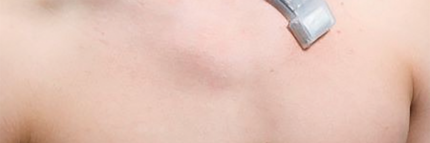
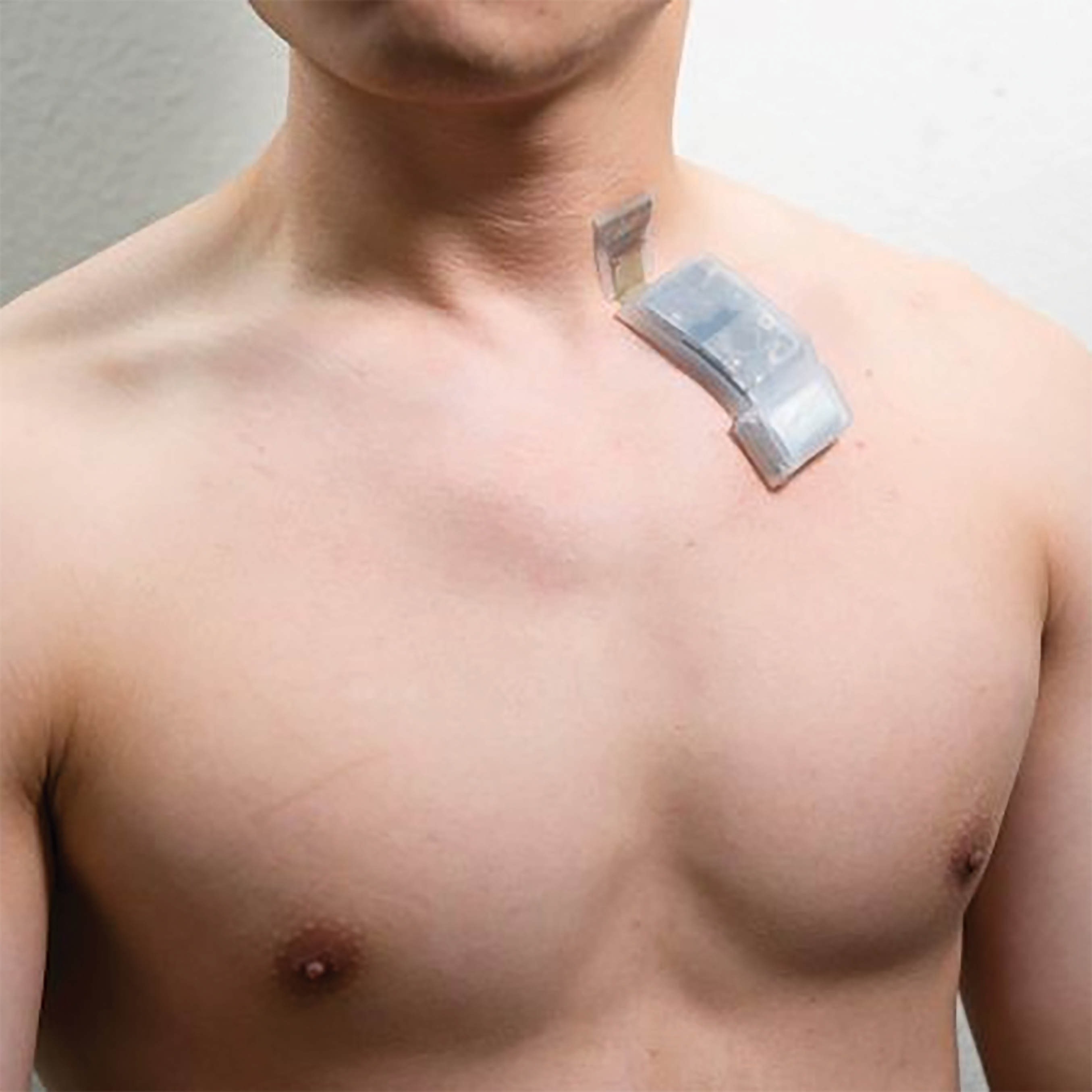
A fully wireless ultrasound patch that can continuously track critical vital signals such as heart rate and blood pressure was recently reported in Nature Biotechnology. The patch, which can capture detailed medical information and wirelessly transmit the data to a smart device (such as a laptop or smartphone), could represent a major step forward in at-home health care technology.
“The true impact of wearable ultrasound patches has yet to be fully realised, as previously described iterations aren’t wireless and limit the users’ ability to go about their daily lives,” noted Dr Randy King, a program director in the Division of Applied Science & Technology at NIBIB. “The technology described here represents necessary and essential progress in the wearable ultrasound space, potentially unlocking the promise of remote ultrasound monitoring for any number of health conditions.”
Ultrasound, which uses sound waves and their resulting echoes to image tissues inside the body, has traditionally been limited to the clinic. The ultrasound patch technology in this study was pioneered by Dr Sheng Xu, an associate professor and Jacobs Faculty Scholar at the University of California San Diego (UC San Diego). His team has previously reported wearable ultrasound patches with similar transducers. The real advancement in this latest study is the wireless capability of the patch.
“The key element of this study is the design of the ultrasound circuit,” said Xu. “In our previous patches, the ultrasound probe was connected to a flexible cable for power and data transmission. In this patch, the cables are replaced with a wearable circuit, which can pre-process and wirelessly transmit the ultrasound data to a back-end station for further analysis.” The ultrasound system is composed of a probe, a circuit, and a battery. As the current system was designed with a focus on cardiovascular health, the ultrasound probe was typically placed on the carotid artery in this study. The probe is attached to a flexible circuit, which activates the ultrasound transducers, collects the ultrasound echoes, amplifies and filters these echoes, and transmits the digitised signal to a terminal device. The entire system is powered by a commercial rechargeable lithium polymer battery.
“We developed a machine learning algorithm to coordinate with the circuit to automatically process the ultrasound signals and continuously track the carotid artery, which allows us to obtain ultrasound information even when the wearer of the patch is moving,” explained first author Dr Muyang Lin, a PhD candidate in the Xu laboratory. “This automatic tracking algorithm provides unprecedented opportunities for medical ultrasonography and exercise physiology.”
To assess its generalizability, the researchers cross-validated their machine learning model among ten healthy subjects representing three distinct racial groups. Using a machine learning technique called domain adaptation, the researchers found that a model trained on data from one participant wearing the patch could be successfully adapted for the other participants. With minimal model retraining, the patch could track the pulsations of the carotid artery with high accuracy, allowing for measurements like blood pressure, arterial stiffness, and cardiac output. Continual monitoring of these measurements among high-risk populations could provide advance warning of heart failure.
“Validating our patch in a larger population is a crucial next step,” said Lin. “We are working on validating our sensor against existing medical devices.”
While the device was mainly evaluated on its ability to monitor cardiovascular functions, the researchers also demonstrated that the patch can be applied to the abdomen for diaphragm monitoring or to limbs for peripheral artery monitoring. “The system holds the potential to perform measurements at multiple spots in the body, and we can easily tailor the probe design to fit diverse tissue monitoring requirements,” said Xu.
“With this kind of device, we hope to blur the boundary between at-home care and in-hospital diagnosis,” said Lin. “We foresee a future where diagnoses can occur anytime and anywhere, enabled by wireless devices like these.”
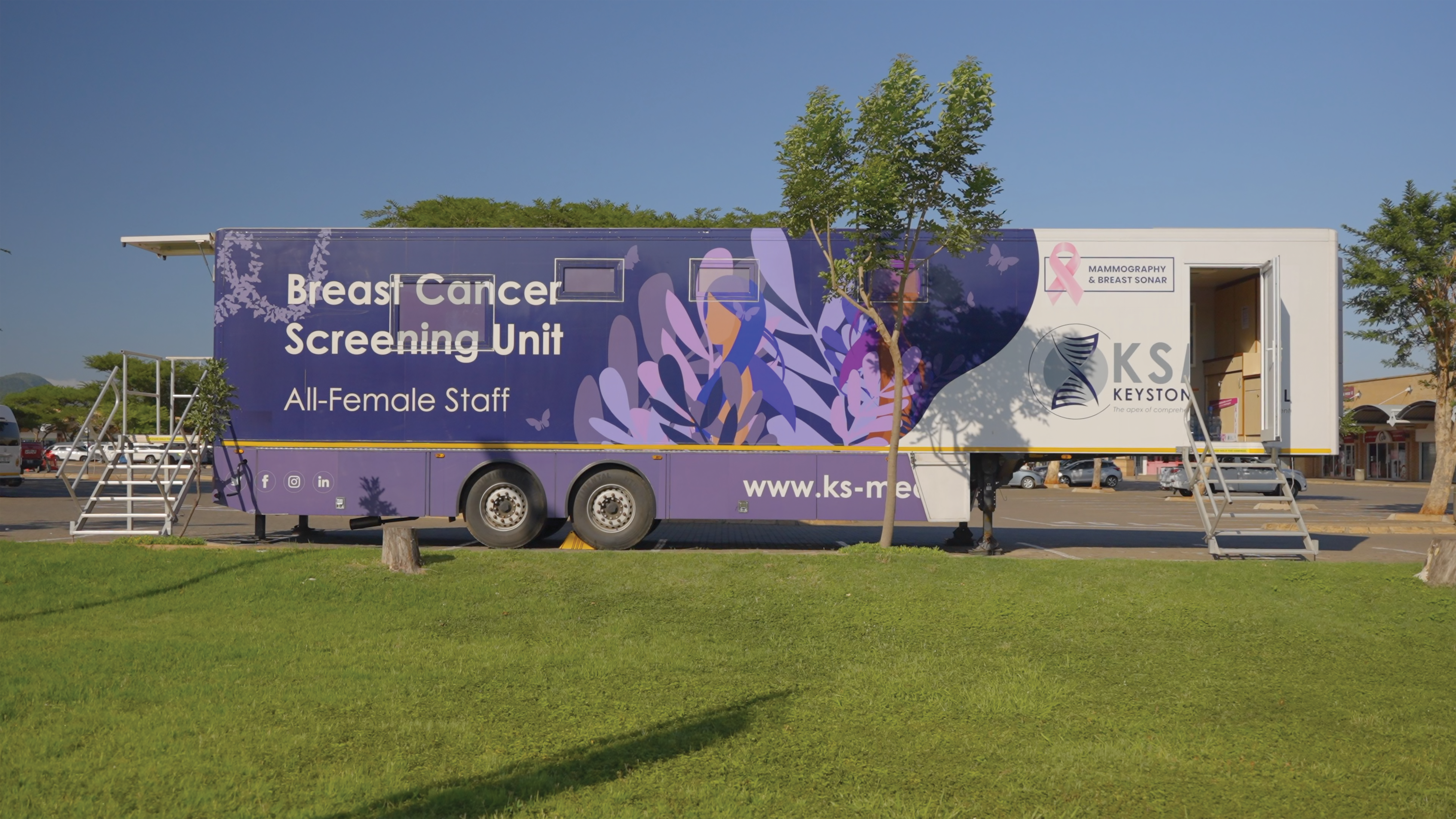
MAJOR DRIVE TO ROLL OUT SCREENING UNITS TO HELP FIGHT BREAST CANCER INRURAL AREAS
Save: Lives can be saved if proactive stance is taken to reduce the treatment burden of breast cancer
By Thuli Zungu
Keystone Medical Group and the Non-Government Organization ‘Screen Her Save Her’ have rolled out a mobile mammography screening unit in three rural areas in Eastern and Western Cape.
Mammography is a technique using X-rays to diagnose and locate tumours of the breasts.
With the mobile mammography screening unit, these organizations aim to provide women with a safe, out of hospital environment for reliable screening and will be targeting women who typically need to travel long distances to access this level of screening.
Rosa-Marie Cox-Cronje, director of Screen Her Save Her, says the incidence rate of breast cancer among South African women currently accounts for over 23% of all cancer cases. Evidence suggests much of the pain, suffering and loss of life associated with this debilitating disease, can be prevented through early detection. Cox-Cronje says statistics collated by the South African government suggests that about 90% of breast cancer patients survive many years after diagnosis when the cancer is detected during its early stages.
“By taking a proactive stance in the fight against breast cancer, we can greatly reduce the related treatment burden and most importantly, save lives. With the mobile mammography screening unit, we aim to provide women with a safe, out of hospital environment for reliable screening. As in previous years, we will be targeting women who typically need to travel long distances to access this level of screening.”
Mammograms and breast ultrasounds are painless examinations that take around 30 minutes to complete. Cox-Cronje says these measures, supplemented by regular self-examinations, have been found to drastically increase the likelihood of detecting breast cancer in its earlier stages and initiating the appropriate treatment.
“Initiatives that support women in accessing the right amount and level of medical care as a way of avoiding the devastating impact of breast cancer, are an integral step towards building a healthier nation’’. They are looking forward to welcoming women within the two rural areas and providing them with the insight and guidance they need to live long, fulfilled lives.
The mobile breast screening unit will be located at the following destinations: · Kathu until 21 July 2023 · Upington from 24 July to 4 August 2023 · Langebaan from 7 to 16 August 2023. Screenings will be made available to both public patients and those with medical aid policies.
A limited number of screenings for women who cannot afford the cost involved, will be offered by SCREEN HER SAVE HER, on a first-come, firstserved basis. Appointments can be pre-booked online via this link: https://calendly.com/ks-med or via the telephone line: 087 055 0587 (option 5).